FEMORAL SHAFT FRACTURES – PEDİATRİC

Prepared by Oguzhan BULUT

SUMMARY

  Femoral shaft fractures are among the most prevalent pediatric orthopedic injuries and stand as the primary cause of hospital admission for orthopedic trauma in children.

  While treatment remains a subject of debate, there is a modern transition from traditional non-operative methods toward surgical stabilization due to advancements in fixation techniques and bone biology.

  Traditional reliance on non-operative care stems from the fact that children possess high osteogenic potential, leading to rapid fracture healing and generally favorable outcomes.

  Management is highly tailored to the patient’s age and fracture morphology, ranging from a Pavlik harness or spica cast to operative fixation.

ETIOLOGY and EPIDEMIOLOGY

  • %2 of all pediatric fractures.
  • It shows a bimodal distribution, divided into 2-4 year olds and adolescent age groups.
  • Males more commonly affected 2.6 / 1 male to female.

Mechanism of injury;

  • Non-Ambulatory Children: Child abuse is the leading cause of femoral fractures before walking age, accounting for 70% to 80% of cases.
  • Toddlers (1–4 years): Approximately 30% of femoral shaft fractures in this age group are attributed to abuse.
  • Children < 10 years: Accidental falls are the most common overall cause.
  • Children > 10 years: Motor vehicle accidents represent the most frequent cause of injury.

Clinical Red Flags and Risks

  • Indicators of Suspicion: Beyond age, factors such as being a first-born child, pre-existing brain damage, bilateral fractures, specific fracture patterns (subtrochanteric or distal metaphyseal beak fractures), and a delay in seeking medical help should raise concern.
  • Safety Implications: Diligent analysis is mandatory; returning a child to an abusive home results in a 50% risk of repeat abuse and a 10% mortality rate.

Differential Diagnosis: Pathologic Fractures

  • Bone Lesions: Radiographs must be evaluated for conditions that predispose bones to fracture, most commonly benign lesions like aneurysmal bone cysts, unicameral bone cysts, and non-ossifying fibromas (NOF).
  • Malignancies: Though less common, Ewing’s sarcoma and metastatic disease must be considered.
  • Systemic Conditions: Medical investigations should rule out osteogenesis imperfecta, neuromuscular disorders, and secondary osteopenia.

ANATOMY

Bone Development and Morphology

  • Ossification Process: Growth begins through enchondral ossification, which leads to the formation of a medullary cavity characterized by peripheral calcification and central vascularization.
  • Bone Maturation: Woven bone is produced during this initial ossification and persists for the first 18 months of life. It is eventually replaced by lamellar bone, with longitudinal and peripheral growth continuing until skeletal maturity.
  • Structural Changes: As a child ages, both the cortical diameter and the cortical thickness of the femoral shaft progressively increase.

Anatomical Landmarks

  • Isthmus: This is identified as the narrowest portion of the femoral shaft, which is a critical consideration for intramedullary fixation.
  • Femoral Bow: Clinicians should always evaluate the anterior bow of the femur, as this curvature affects implant selection and alignment.

Muscles

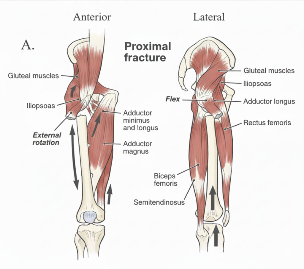
  Proximal Fragment: The iliopsoas muscle exerts a specific deforming force on the proximal bone segment, pulling it into flexion and external rotation.

  Distal Fragment: The adductor muscles act primarily on the distal bone segment, creating forces that result in shortening and varus (inward) angulation of the limb

PHYSICAL EXAMINATION

  • The patient has localized tenderness and swelling and usually has deformity with associated shortening and obvious crepitus on palpation.
  • Neuravascular examination should be carefully noted.
  • Skin areas of spine and pelvis needs to thoroughly investigated for ecchymosis.
  • Every extremity must be carefully inspected and palpated to avoid missing injuries, whch can become difficult to treat if detected late.
  • This is especially important in the head-injured patient who is unable to communicate symptoms.
  • Repated examinations are a necesserary part of the evaluation in this situation.

RADIOLOGICAL EXAMINATION

  • Standart AP and Lateral of entire femur including knee and pelvis are necessary.
  • Poor-quality radiographs are unacceptable and study must be repated.
  • The x-rays should be evaluated for fracture configuration, degree of comminution, displacement, angulation and degree of shortening.
  • CT and MRI are usefull for pathological fractures or stres fractures.
  • Plain radiographs are usually all that is needed.

CLASSIFICATION

  • The classification of femoral shaft fractures based on radiographic examination and the condition of the soft tissue envelope.
  • Closed or open fracture.
  • Location: Proximal, middle, distal third.
  • Configuration: Transverse, oblique, spiral.
  • Communation and shortening; >3 cm shortening is unacceptable.

TREATMENT

  • The age and size of the child are the most important factors in deciding which treatment modality is most appropriate.
Patient Age GroupFracture CharacteristicsPrimary Treatment Options
Infants (< 6 months)All fracture patternsPavlik harness or Early spica casting
Young Children (6 months – 5 years)Stable: Simple patternsEarly spica casting
Complex: Unstable patterns, polytrauma, or open fracturesExternal fixation or Traction followed by delayed spica casting
School Age (5 – 11 years)Length Stable: Weight < 49kg (100 lbs)Flexible titanium nails (TENS)
Length Unstable: Comminuted/spiral patterns, proximal/distal locations, or regardless of weightORIF with submuscular bridge plating, Stainless steel Enders nails, or External fixation
Adolescents (> 11 years)Standard: Weight > 49kg (100 lbs)Antegrade rigid intramedullary nail fixation
Complex: Highly comminuted, or proximal/distal metaphyseal involvementORIF with submuscular bridge plating
  • 0 to 6 months: immediate application of Pavlik harness results in an excellent outcome with time to union about 5 weeks. Excessive hip flexion in the presence of a swollen thigh may lead to a femoral nerve palsy and therefore weekly evaluations of quadriceps function should be performed during treatment.
  • 6 months to 5 years: when shortening of the fracture is limited less than 3 cm with a stable pattern, prefer to treat with closed reduction and spica casting.Skin or sceletal traction is requried when excess shortening or angulation is present. In the multiple injured patient, immediatestable fixation is often required    and is bes accomplished with external fixation or plate fixation.
  • 6 to 10 years :  Treated with closed or open reduction and stabilized with flexible rods especially when a stable transvers fracture pattern is present.
  • 11 years to skeletal maturity: Intramedullary rodding is agan is excellent choice in a stable fracture pattern . In this age of group the rigid locked intramedullary rod is preferred in some centers for a stable fracture pattern, but it must be placed with the proximal starting point at or jusst distal to the greater trochanter to minimize the risk of AVN.

TECHNIQUES:

1. Pavlik Harness

This method is typically reserved for very young infants (usually under 6 months old).

  • Technique:  This approach is advantageous because it avoids the need for anesthesia.
  • Complications:  Excessive hip flexion, especially in the presence of a swollen thigh, can compress the femoral nerve.
    • This nerve compression is identified by decreased quadriceps function.

2. Immediate Spica Casting

Spica casting is a common definitive treatment for toddlers and young children.

  • Technique:
    • Unlike the harness, this is applied with reduction under sedation or with general anesthesia.
    • It can be applied as a single-leg spica or one-and-one-half spica (used specifically to control rotation).
    • Note: A distal femoral buckle fracture may be treated with a long leg cast alone rather than a full spica.
  • Positioning & Alignment:
    • Hips should be flexed 60-90° with approximately 30° of abduction.
    • External rotation is typically required to correct rotational deformities.
    • The cast is molded into recurvatum and valgus; this counteracts muscular forces that naturally pull the fracture into procurvatum and varus.
    • Specific molds along the distal femoral condyles and buttocks help maintain the reduction.

  • Regardless of the method used, the following clinical targets apply:
  • Acceptable Limits: These are generally based on the patient’s age (due to remodeling potential).
  • Primary Goals:
    • Coronal plane: < 10° deformity.
    • Sagittal plane: < 20° deformity.
    • Shortening: No more than 2cm.
    • Rotation: < 10° of rotational malalignment.
  • Logistics: A special car seat is sometimes needed for transport, though a regular car seat can often be used if a single-leg spica was applied.

3.External Fixation

Indications

In modern practice, external fixators are primarily indicated for complex scenarios where internal fixation or casting is not ideal. These include:

  • Open fractures or fractures with severe disruption of the soft tissue envelope (including severe burns).
  • Multiple trauma (polytrauma) or cases involving arterial injury.
  • Unstable fracture patterns or instances where non-operative methods have failed.

Surgical Technique & Application

  • Placement: The fixator is applied laterally to avoid disruption and scarring of the quadriceps muscle.
  • Duration: The fixator is generally left in place for 10 to 16 weeks until solid bony union has been achieved.
  • Mobility: Full weight-bearing is permitted as tolerated while the fixator is in place.

Complications & Risks

  • Refracture: There is an increased risk of refracture after removal, which is a particular concern in oblique fracture patterns.
  • Pin-Track Care: (Note: While not in your original list, this is a standard clinical consideration for external fixators to prevent infection).

4. Submusculer Plate Fixation

Surgical Approach & Technique

  • Approach: A laterally based incision and plating method designed for minimal disruption of the soft tissue envelope.
  • Incision Placement: Small proximal and distal incisions are made; the plate is then placed between the periosteum and the vastus lateralis on the lateral side of the femur.
  • Reduction: The fracture is provisionally reduced using closed or percutaneous techniques before the plate is fixed.

Instrumentation & Hardware

  • Plate Selection: Typically uses a 12–16 hole, 4.5mm narrow LC-DCP (Limited Contact Dynamic Compression Plate).
  • Fixation: Securement usually requires 3 screws proximal and 3 screws distal to the fracture site.
  • Contouring: The plate may need to be manually bent to accommodate the natural curvature of the femur, though pre-contoured femur plates are also an option.

Outcomes & Complications

  • Clinical Outcomes: This method generally shows favorable results regarding time to union, early weight-bearing, reduced hardware irritation, and lower incidence of a postoperative limp.
  • Potential Complications:
    • Need for hardware removal.
    • Risk of refracture following the removal of hardware.

A quick technical note

When you mention the “natural bend of the femur,” surgeons often refer to this as the anterior bowing of the femoral shaft. If you are using a straight LC-DCP, ensuring that longitudinal contouring matches this bow is critical to avoid “propped” reductions or malalignment.

Could you rearrange these sentences and give them back to me?

5.Flexible Intramedullary Nail (Titanium Elastic Nail) 

All-Distal Approach

  • Incision: 2cm incisions are made medially and laterally at the level of the distal physis.
  • Procedure: Soft tissue is spread with a hemostat to reach the starting point, located approximately 2cm proximalto the physis.

Distal and Proximal Approach

  • Incision: A 2cm incision is made laterally at the level of the distal physis, combined with a 2cm incision proximally at the greater trochanteric apophysis.

Instrumentation & Sizing

  • Nail Sizing Formula: Nail size is determined by multiplying the width of the femoral canal isthmus by .
  • Canal Fill: The surgical goal is to achieve 80% canal fill (e.g., two nails each filling 40% of the narrowest part of the canal).

Outcomes

  • General Results: Outcomes are generally excellent with high patient satisfaction.
  • Healing Time: Time to union is typically 10–12 weeks.
  • Hardware Removal: Removal of the nails is generally performed at the 1-year mark.

Complications

Insertion Site Pain

  • Frequency: This is the most common complication, occurring in up to 40% of patients, specifically near the knee.
  • Prevention: To minimize irritation, it is recommended that nail protrusion remains < 25mm with a minimal bend outside the femur.

Malunion & Patient Factors

  • Risk Factors: There is an increased rate of complications in patients who are  11 years old or weigh  kg.
  • Fracture Characteristics: Increased rates of malunion are seen with:
    • Comminuted fractures.
    • Highly shortened fractures.
    • Fractures located in the very proximal or distal segments of the bone.

6.Antegrade Rigid Intramedullary Nail Fixation

Surgical Approach & Entry Points

  • Preferred Techniques:
    • Lateral Entry Nail
    • Trochanteric Entry Nail (Starting point at the tip of the greater trochanter).
  • Entry Point Avoidance: Piriformis entry must be avoided due to the high risk of injuring the vascular supply to the femoral head.
  • Soft Tissue Dissection: * A lateral incision is made proximal to the greater trochanter.
    • Sharp dissection or electrocautery is used through the fascia lata to reach the starting point at the tip of the greater trochanter.
  • Reduction: The fracture is managed via either closed or open reduction techniques.

Instrumentation & Insertion

  • Procedure: Once the fracture is reduced, follow standard steps to insert the intramedullary nail.
  • Safety Precaution: Exercise extreme caution to ensure the nail does not cross the distal physis, preserving the growth plate.

Complications

Osteonecrosis (AVN) Risks

  • Piriformis Entry: In patients with open proximal physes, the risk of osteonecrosis is 1–2%.
  • Alternative Entry: The exact risk of osteonecrosis associated with greater trochanteric and lateral entry nails is currently unknown.

Proximal Femur Deformities

Greater trochanteric insertions can lead to secondary deformities, including:

  • Premature fusion of the greater trochanter apophysis.
  • Coxa valga and subsequent hip subluxation.
  • Narrowing of the femoral neck.

Outcomes

  • Clinical Success: This method yields favorable outcomes, particularly in the adolescent population.
  • Alignment: There is a significantly decreased risk of angular malunion compared to other fixation methods in this age group.

COMPLICATIONS

1. Limb-Length Inequality (LLI)

  • Overview: LLI is the most common complication following a femoral shaft fracture in children. It can result from either shortening at the fracture site or accelerated growth (overgrowth) in the involved femur.
  • Timeline: Typically manifests within 2 years of the injury.
  • Clinical Thresholds: * Shortening is generally considered acceptable if less than 2–3 cm, as overgrowth is anticipated in younger patients.
    • Shortening becomes symptomatic if it exceeds the 2–3 cm threshold.

2. Osteonecrosis & Vascular Risks

  • Contraindication: Femoral nailing through the piriformis fossa is strictly contraindicated in adolescents with open physes due to the high risk of osteonecrosis of the femoral head.
  • Vascular Anatomy: The primary blood supply to the femoral head is the deep branch of the medial femoral circumflex artery. This artery branches into the superior retinacular vessels, which are highly vulnerable as they lie in close proximity to the piriformis fossa.
  • Entry Point Risks: While most associated with the piriformis start, osteonecrosis has been reported with both piriformis and greater trochanter entry nails.

3. Nonunion & Malunion

  • Fixation Type: There is a higher risk of nonunion or malunion when using load-bearing devices, such as external fixators or submuscular plates.
  • Patient Factors (Flexible Nails): When using flexible intramedullary nails, the risk of malunion or nonunion increases significantly in patients who are:
    • Over 11 years old.
    • Over 49 kg (108 lb) in body weight.
  • The typical deformity is varus + flexion of the distal fragment, remodeling is greatest in the sagittal plane, rotational malalignment does not remodel.
  • Nearly 50% of fractures treated with flexible nails have 15 degrees of malalignment

References

Flynn JM, Skaggs DL, Sponseller PD, et al. The treatment of pediatric femoral shaft fractures. J Am Acad Orthop Surg. 2015;23(11):e1–e11.

Beaty JH, Kasser JR. Rockwood and Wilkins’ Fractures in Children. 8th ed. Wolters Kluwer; 2015.

Kocher MS, Sink EL, Blasier RD, et al. Treatment of pediatric diaphyseal femur fractures. J Am Acad Orthop Surg. 2009;17(11):718–725.

American Academy of Orthopaedic Surgeons. Clinical Practice Guideline: Pediatric Diaphyseal Femur Fractures. AAOS; 2020.

Flynn JM, Hresko T, Reynolds RA, et al. Titanium elastic nails for pediatric femur fractures. J Pediatr Orthop. 2001;21(1):4–8.

Ligier JN, Metaizeau JP, Prévot J, Lascombes P. Elastic stable intramedullary nailing of femoral shaft fractures in children. J Bone Joint Surg Br. 1988;70-B:74–77.

Luhmann SJ, Schoenecker PL. Complications of titanium elastic nails for pediatric femoral shaft fractures. J Pediatr Orthop. 2003;23(4):443–447.

Kocher MS, Kasser JR. Orthopaedic aspects of child abuse. J Am Acad Orthop Surg. 2000;8(1):10–20.

Leave a Reply

Your email address will not be published. Required fields are marked *