PEDIATRIC PROXIMAL FEMORAL FRACTURES

Prepared by Dr. Ahmet Mucteba YILDIRIM

Overview

  • Epidemiology: These are high-energy injuries; 30% are associated with major trauma (pelvic or intra-abdominal).
  • Fracture Mechanism: Typically results from high-velocity accidents (falls from height, motor vehicle accidents). In infants <12 months, child abuse must be ruled out.
  • Risk of Complications: Despite advancements in implant technology, the risk of complications such as AVN (Avascular Necrosis), premature physeal arrest, and coxa vara remains higher compared to other injuries.
  • Pathologic Fractures: Consider underlying lesions such as simple bone cysts, fibrous dysplasia, or spina bifida.

Clinical Presentation

  • Symptoms: Acute pain, inability to bear weight, and deformity.
  • Delayed Diagnosis: In non-displaced fractures, diagnosis may be missed; the patient may only present with a persistent limp after a minor sports injury.

Physical Examination

  • Observation: The affected limb is usually shortened and externally rotated in displaced fractures.
  • Palpation & ROM: Global limitation of hip movements due to pain.

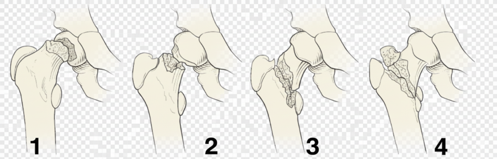
Delbet Classification and Treatment Approaches

The incidence of AVN decreases as the fracture line moves distally.

Type 1: Transphyseal Fractures (Highest AVN Risk)

Type 1 epiphysiolysis. Unlike SCFE, it involves higher energy and no prodromal period. It may be seen in newborns with breech presentation and be misdiagnosed as congenital hip dislocation. It may be seen during reduction of hip dislocation.

Treatment: Non-repetitive closed reduction followed by percutaneous fixation is performed. If reduction cannot be achieved, open reduction (using Moore or Smith-Petersen approaches) is executed.

Implant Selection: K-wires for children under 4 years; 4.0 mm screws for ages 4–7; 5.0 mm or 6.5 mm cannulated screws for those over 7 years.

To reduce the risk of AVN, hematoma aspiration and pelvic-pedal casting (hip spica) are recommended.

Type 2: Transcervical Fractures (Most Common – 50%)

Fixation is recommended even in non-displaced cases to avoid complications. Forceful closed reduction should be avoided due to AVN risk; open reduction via anterior or anterolateral approach should be performed if necessary.

In adolescents, at least two screws should be placed to provide compression. If the Pauwels angle is high or the fracture is close to the head, crossing the physis should not be avoided to ensure union. 6–12 weeks of pelvic-pedal casting follows treatment.

Type 3: Cervicotrochanteric Fractures

While the incidence of AVN is lower than in Type 2, the risk of coxa vara is similar. Surgical treatment is indicated for all displaced fractures and for non-displaced fractures in children over 6 years old. Locking plates can be used to prevent varus collapse; otherwise, cannulated screws with a hip-spica cast are applied.

Type 4: Pertrochanteric Fractures: This group has the lowest observed risk of AVN.

Complications and Management

AVN (Avascular Necrosis): Incidence rates for Type 1, 2, 3, and 4 are 38%, 28%, 18%, and 5%, respectively. Risk increases in Type 1–2 fractures, patients over 12 years old, and cases where intervention occurs after 24 hours. Capsular decompression is recommended to reduce this risk. Type 1 (total AVN) is the most common and has the worst prognosis.

Coxa Vara: Observed in 10–32% of cases. It results from inadequate perioperative reduction, loss of postoperative reduction (in groups without hip-spica), or premature physeal arrest. Pauwels valgus osteotomy is recommended when the neck-shaft angle falls below 110 degrees.

Non-Union: Occurs in 6–12% of cases. Risk increases with inadequate reduction, distraction at the fracture line, or a Pauwels angle >50 degrees. For patients under 10, iliac autografting and compressive screw fixation are recommended; for those over 10, subtrochanteric valgus osteotomies (with DHS or locking plates) are preferred to improve weight distribution.

Premature Physeal Arrest: Since the proximal femoral epiphysis contributes only 15% to overall growth, serious limb length discrepancy (LLD) is usually not seen. Screws penetrating the physis increase this risk.

Pearls & Pitfalls

Pearls (Clinical Tips)
  • Assume high-energy until proven otherwise: These injuries often coexist with major trauma—perform full trauma assessment and screen for associated injuries.
  • Always consider non-accidental trauma in infants (<12 months): Especially low-energy history, inconsistent story, or delayed presentation.
  • AVN risk is highest proximally: Delbet type I–II carry the greatest risk; aim for anatomic reduction and stable fixation.
  • Gentle reduction matters: Avoid repeated/forceful closed reduction attempts—especially in type I–II—because iatrogenic vascular compromise may increase AVN risk.
  • Low threshold for open reduction: If closed reduction is not acceptable on the first attempt, proceed to open reduction (anterior/anterolateral approaches commonly used).
  • Capsular decompression: Consider decompression (hematoma aspiration/capsulotomy) particularly in high-risk patterns (type I–II, displaced fractures) to reduce intracapsular pressure.
  • Fixation strategy should match age and fracture pattern:
    • <4 years: K-wires are commonly used
    • 4–7 years: small cannulated screws (e.g., 4.0 mm)
    • >7 years/adolescents: larger cannulated screws (e.g., 5.0–6.5 mm); typically ≥2 screws for compression and rotational control
  • Do not fear crossing the physis when necessary (selected cases): In adolescents with unstable patterns/high Pauwels angle or very proximal fractures, crossing the physis may be justified to obtain union—document the rationale.
  • Prevent varus collapse: For cervicotrochanteric patterns (type III), consider implants that resist varus (e.g., locking plate constructs) when indicated.
  • Immobilisation is adjunctive, not a substitute for fixation: Hip spica may help protect the repair in younger children, but quality reduction and stable fixation remain primary.

Pitfalls (Common Errors)

  • Missing nondisplaced fractures: A child with persistent limp after minor trauma can still have a proximal femoral fracture—maintain suspicion and obtain appropriate imaging.
  • Repeated reduction attempts: Multiple manipulations increase soft tissue trauma and may worsen perfusion.
  • Accepting a poor reduction: Malreduction increases risk of nonunion, coxa vara, and poor function.
  • Inadequate fixation: Too few screws, poor screw spread, or insufficient purchase can lead to loss of reduction and nonunion.
  • Ignoring varus tendency: Type III fractures are prone to varus; without adequate support, coxa vara can develop.
  • Delayed treatment without escalation: Higher AVN risk is associated with delayed intervention—particularly in high-risk patterns—so avoid unnecessary delays once diagnosis is confirmed.
  • Unclear postoperative plan: Lack of structured follow-up and protection (weight-bearing/immobilisation) can contribute to mechanical failure.
  • Failure to counsel families: AVN and growth-related complications must be discussed early; document informed consent clearly.

Treatment Algorithm (Delbet Classification-Based)

Key principle: AVN risk decreases as the fracture line becomes more distal (Type I highest → Type IV lowest).
Primary goals: early diagnosis, anatomic reduction, stable fixation, and complication prevention.


Step 1 — Initial Assessment

  1. Stabilize and screen (high-energy trauma protocol when applicable).
  2. Rule out abuse in infants and non-ambulatory children.
  3. Obtain imaging:
  • AP pelvis + lateral hip (and additional views as needed)
  • Consider MRI/CT if occult injury suspected (case-dependent)

Step 2 — Classify the Fracture (Delbet I–IV)

  • Type I: Transphyseal
  • Type II: Transcervical
  • Type III: Cervicotrochanteric
  • Type IV: Pertrochanteric

Step 3 — Decide by Displacement (All Types)

A) Nondisplaced

  • Type II: Fixation is generally recommended even if nondisplaced (high complication risk).
  • Type III–IV: Consider fixation based on age and stability; in older children, fixation is commonly preferred.

B) Displaced

  • All types: Reduction + internal fixation is typically indicated.

Step 4 — Reduction Strategy

  • First attempt: Single, gentle closed reduction (avoid forceful maneuvers).
  • If unacceptable reduction: proceed to open reduction (especially Type I–II and unstable patterns).

Step 5 — Fixation Strategy (Practical Guide)

Type I (Transphyseal) — Highest AVN risk

  • Reduction: Non-repetitive closed reduction → if not achieved, open reduction
  • Fixation (age-based examples):
    • <4 years: K-wires
    • 4–7 years: small cannulated screws (e.g., 4.0 mm)
    • 7 years: 5.0–6.5 mm cannulated screws
  • Adjuncts: capsular decompression / hematoma aspiration; hip spica may be considered in younger children

Type II (Transcervical) — Most common (~50%)

  • Fixation recommended even when nondisplaced
  • Avoid forceful reduction; low threshold for open reduction if needed
  • Adolescents: typically place ≥2 screws for compression/rotational control
  • Consider crossing the physis if required for stability/union in very proximal or high-angle fractures (document rationale)
  • Post-op protection: hip spica or restricted weight bearing based on age/stability (often 6–12 weeks in younger patients)

Type III (Cervicotrochanteric)

  • Lower AVN than Type II, but coxa vara risk remains significant
  • Treatment:
    • Displaced: operative fixation
    • Nondisplaced: consider fixation in children >6 years
  • Implants: locking plates can help prevent varus collapse; alternatively cannulated screws + hip spica in selected cases

Type IV (Pertrochanteric) — Lowest AVN risk

  • Usually managed operatively if displaced; stable patterns may be individualized
  • Focus on stable fixation and early functional recovery (age and stability dependent)

Step 6 — Postoperative Follow-up and Complication Surveillance

  • Serial radiographs to monitor union and alignment
  • Monitor for:
    • AVN (early signs may be subtle)
    • Coxa vara (neck–shaft angle trend)
    • Nonunion (pain, lack of progression on imaging)
    • Premature physeal arrest (especially if physis crossed)

Mini Management Notes for Major Complications

AVN
  • Highest in Type I–II; risk increases with older age and delayed intervention
  • Consider capsular decompression in high-risk patterns
  • Prognosis is worst in total head involvement
Coxa Vara
  • Often due to inadequate reduction, loss of reduction, or physeal arrest
  • Consider Pauwels valgus osteotomy when neck–shaft angle <110°
Nonunion
  • Risk factors: inadequate reduction, distraction, high Pauwels angle (>50°)
  • <10 years: iliac autograft + compressive fixation
  • 10 years: subtrochanteric valgus osteotomy (DHS or locking plate) to improve biomechanics
Premature Physeal Arrest
  • Proximal femoral physis contributes ~15% of overall growth → major LLD often limited
  • Risk increases when screws penetrate the physis

References

  1. Rockwood and Wilkins’ Fractures in Children. 9th ed. Philadelphia: Wolters Kluwer; 2019. (Proximal femur fractures chapter)
  2. Beaty JH, Kasser JR (eds). Skeletal Trauma in Children. 6th ed. Philadelphia: Elsevier; 2020. (Femoral neck / proximal femur fractures)
  3. Flynn JM, Skaggs DL, Waters PM (eds). Rockwood and Wilkins’ Fractures in Children / pediatric trauma reference chapters on proximal femoral fractures (latest available edition).
  4. Canale ST, Beaty JH. Campbell’s Operative Orthopaedics. 14th ed. Philadelphia: Elsevier; 2021. (Pediatric hip/femoral neck fractures; fixation strategies and complications)
  5. Delbet P. Fractures of the neck of the femur in children. Rev Chir (Paris). 1907. (Original classification—historical)
  6. Ratliff AHC. Fractures of the neck of the femur in children. J Bone Joint Surg Br. 1962;44-B:528–542. (Classic outcomes/complications including AVN)

Leave a Reply

Your email address will not be published. Required fields are marked *