Fibrous Dysplasia

Prepared by Dr. Erhan OKAY

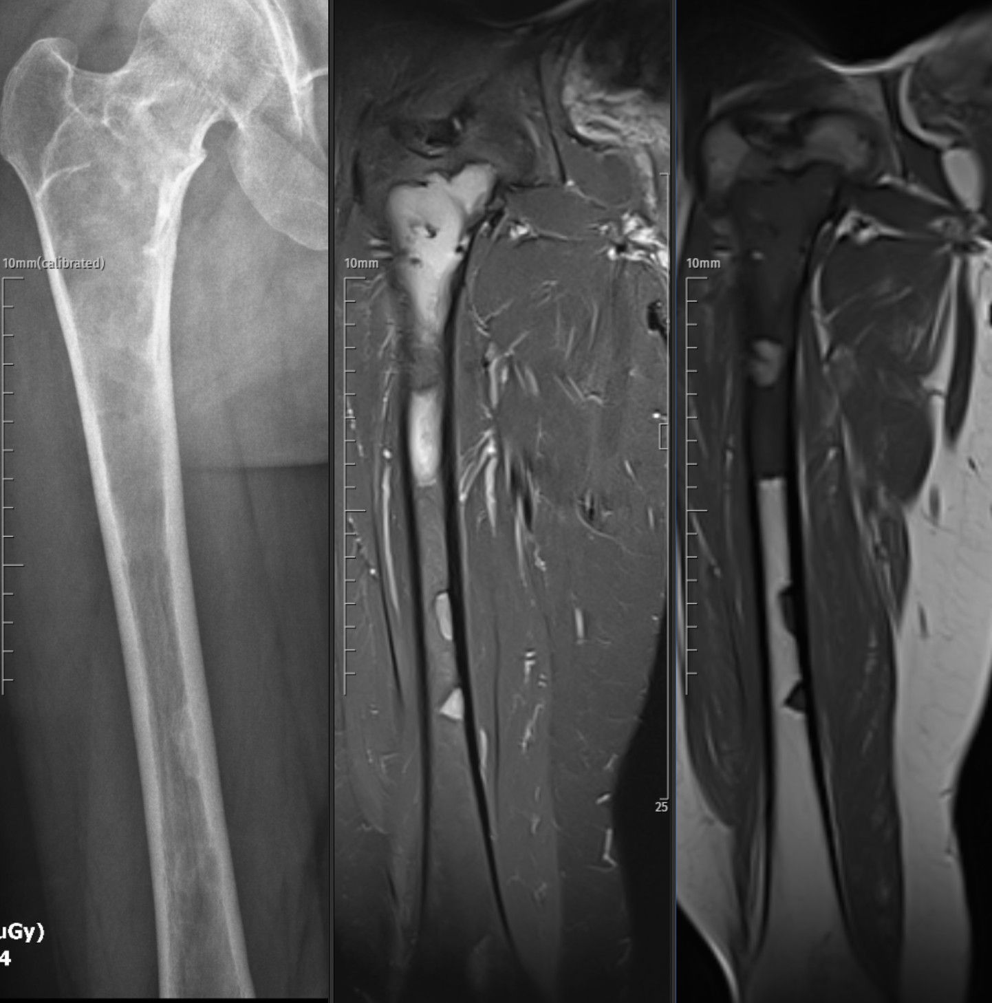
Fibrous dysplasia (FD) is a benign bone disorder characterized by the replacement of normal bone with fibro-osseous tissue, leading to pain, deformity, and fractures. It results from post-zygotic GNAS gene mutations that disrupt osteoblastic differentiation. FD may be monostotic (single bone) or polyostotic, the latter often occurring as part of McCune–Albright syndrome (MAS). Radiologically, it presents with a ground-glass appearance and possible deformities such as the “shepherd’s crook” in the proximal femur. Treatment is primarily symptomatic, involving bisphosphonates for pain control and surgery for deformity or fracture correction. Although benign, the disease may progress during growth and stabilize in adulthood, requiring periodic follow-up for skeletal deformity and functional assessment.

Pathophysiology

  • Caused by post-zygotic mutations in the GNAS gene.
  • Leads to defective osteoblastic differentiation and formation of immature woven bone mixed with fibrous tissue.
  • Two clinical forms:
    Monostotic FD: Involves a single bone.
    Polyostotic FD: Affects multiple bones and may be associated with McCune–Albright syndrome (endocrine abnormalities + café-au-lait spots).

 Clinical Presentation & Imaging

  • Symptoms: Bone pain, deformity, limp, or pathological fracture.
  • Typical signs: “Ground-glass” appearance on radiographs, cortical thinning, and possible cystic changes.
  • Characteristic deformity: Shepherd’s crook in proximal femur due to repeated microfractures.
  • CT scan: Useful for evaluating lesion extent and surgical planning.
  • MRI: May show low-to-intermediate T1 and high T2 signal intensities.

Differential Diagnosis

  • Monostotic FD: Simple bone cyst, osteofibrous dysplasia, osteoblastoma, hemangioma, Paget’s disease.
  • Polyostotic FD: Hyperparathyroidism, enchondromatosis, neurofibromatosis, eosinophilic granuloma.Fibrous dysplasia

Treatment

Medical Management

  • Pain control: NSAIDs for symptomatic relief.
  • Bisphosphonates (Pamidronate): 0.5–1 mg/kg/day IV for 2–3 days every 6–12 months.
    Reduces pain and bone turnover, but does not halt disease progression.
  • Experimental therapies: Calcitonin and other bone-modulating agents under investigation.

Surgical Management

  • Indicated for:
    Structural deformity or pathological fractures.
    Progressive or symptomatic lesions.
  • Procedures: Curettage, bone grafting, internal fixation (e.g., intramedullary nailing), corrective osteotomies.
  • Risks: Recurrence and infection, though outcomes are generally favorable.

Prognosis

  • Usually benign and stabilizes after skeletal maturity.
  • Polyostotic cases may require annual follow-up for deformity monitoring.
  • Disease activity typically declines after adolescence.
  • Functional disability may occur in cases with extensive skeletal involvement.

 References

  1. Hartley I, Zhadina M, Collins MT, Boyce AM. Fibrous Dysplasia of Bone and McCune-Albright Syndrome: A Bench to Bedside Review. Calcif Tissue Int. 2019;104(5):517–529. doi:10.1007/s00223-019-00550-z.
  2. Fitzpatrick KA, Taljanovic MS, Speer DP, et al. Imaging Findings of Fibrous Dysplasia with Histopathologic and Intraoperative Correlation. AJR Am J Roentgenol. 2004;182(6):1389–1398. doi:10.2214/ajr.182.6.1821389.
  3. Riddle ND, Bui MM. Fibrous Dysplasia. Arch Pathol Lab Med. 2013;137(1):134–138. doi:10.5858/arpa.2012.0013-RS.
  4. Lala R, Matarazzo P, Bertelloni S, et al. Pamidronate Treatment of Bone Fibrous Dysplasia in Children with McCune-Albright Syndrome. Acta Paediatr. 2000;89(2):188–193. doi:10.1080/080352500750028816.
  5. Hart ES, Kelly MH, Brillante B, et al. Onset, Progression, and Plateau of Skeletal Lesions in Fibrous Dysplasia and the Relationship to Functional Outcome. J Bone Miner Res. 2007;22(9):1468–1474. doi:10.1359/jbmr.070511.

Plain radiograph and coronal CT image of the leg show a well-circumscribed intramedullary expansile lesion in the proximal fibula with a ground-glass matrix and smooth cortical thinning. No cortical breakthrough or periosteal reaction is present. The imaging features are consistent with monostotic fibrous dysplasia.

Radiograph and coronal MRI images of the proximal femur demonstrate a well-defined intramedullary expansile lesion with a ground-glass matrix and cortical thinning without periosteal reaction or soft-tissue extension. On MRI, the lesion shows heterogeneous low-to-intermediate signal on T1-weighted and high signal on fat-suppressed proton-density images, consistent with a fibrous dysplasia involving the proximal femoral shaft.

AspectDetails
NatureBenign fibro-osseous lesion replacing normal bone with fibrous tissue
Genetic BasisGNAS mutation causing defective osteoblastic differentiation
FormsMonostotic (single bone) and Polyostotic (multiple bones, often with McCune–Albright syndrome)
Common SitesFemur, tibia, ribs, craniofacial bones
Characteristic Imaging“Ground-glass” matrix, cortical thinning, shepherd’s crook deformity (proximal femur)
SymptomsBone pain, deformity, limp, or pathologic fracture
TreatmentPain control (NSAIDs), bisphosphonates for bone turnover reduction, surgery for deformity/fracture
PrognosisBenign course; stabilizes after skeletal maturity; annual follow-up for polyostotic cases

Leave a Reply

Your email address will not be published. Required fields are marked *