Prepared by Dr. Enes KANAY
Unicameral bone cyst (UBC) is a benign, fluid-filled intramedullary lesion typically located in the metaphysis or diaphysis of long bones in children and adolescents. It is usually unilocular and adjacent to the cortex. Pathological fracture is the most common presentation.
1.Associated Conditions
UBC may coexist with other lesions:
2. Epidemiology
3. Pathogenesis
The exact etiology is unclear; proposed mechanisms include intramedullary circulation disturbance and venous obstruction, leading to increased intramedullary pressure and cyst formation. Persistent fluid communication with the growth plate is seen in some cases.
4. Clinical Features
5. Imaging
Simple bone cysts typically appear as well-defined, centrally located, intramedullary lucent lesions within the metaphysis of long bones, most often the proximal humerus or femur. They demonstrate a narrow zone of transition, thin sclerotic margins, and may cause mild endosteal expansion or cortical thinning without cortical destruction or periosteal reaction.
On plain radiographs,they are usually unilocular, although pseudotrabeculation may occasionally be seen. In the presence of a fracture, a fallen fragment sign or rising bubble sign may be visible. CT confirms the cystic nature and extent of the lesion but adds little diagnostic advantage.
On MRI,simple bone cysts show low signal intensity on T1-weighted and high signal on T2-weighted sequences, reflecting their fluid content. Post-contrast images demonstrate only thin peripheral enhancement corresponding to the cyst wall, without solid or nodular components. There is typically no marrow or soft-tissue edema, helping distinguish SBCs from more aggressive cystic or neoplastic processes.
6. Histology
Gross:
Microscopic:
Differential Diagnosis: Aneurysmal bone cyst, intraosseous ganglion, fibrous dysplasia
7. Treatment and Natural History

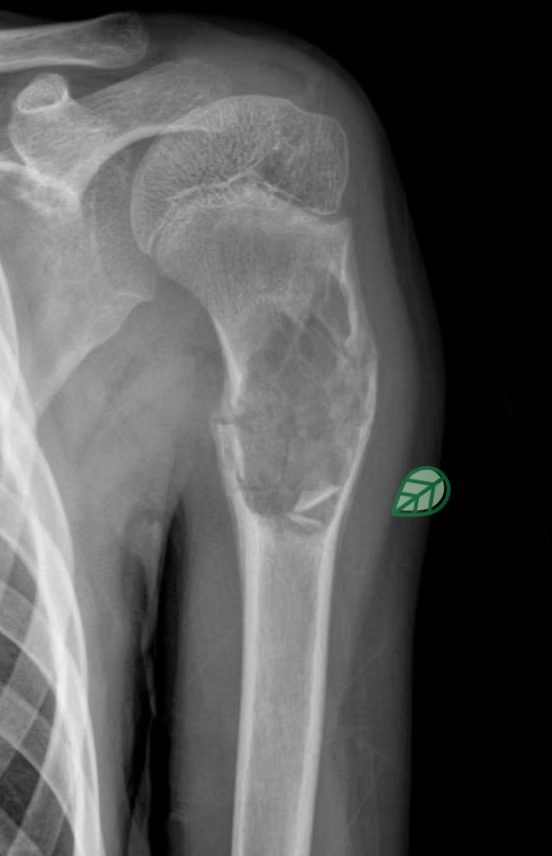
Fallen leaf sign on Xray