Prepared by Dr. Alper DUNKI
Chondroblastoma is a rare, epiphyseal, benign bone tumor that exhibits locally aggressive behavior.
It primarily affects skeletally immature individuals, most commonly males in their second decade of life.
Most frequent locations include the distal femur, proximal tibia, proximal humerus, and less commonly the hip or calcaneus.
Clinical Presentation
Patients typically present with:
Imaging Features
Chondroblastomas typically present as well-circumscribed, lobulated lytic lesions located in the epiphysis or apophysis of long bones, most often around the knee or proximal humerus. On radiographs, they demonstrate geographic bone destruction with a thin sclerotic margin and may contain subtle internal calcifications reflecting a chondroid matrix. CT better delineates these calcifications and cortical integrity.
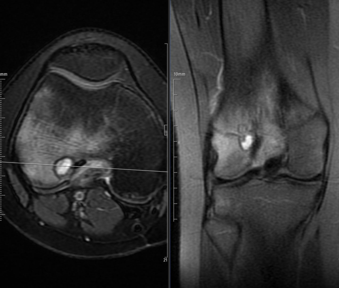
On MRI, chondroblastomas usually appear heterogeneous, with intermediate signal intensity on T1-weighted and variable high signal on T2-weighted or fat-suppressed sequences, often surrounded by bone marrow and soft-tissue edema. A thin hypointense rim corresponding to reactive sclerosis is frequently seen. Post-contrast images show heterogeneous enhancement of the solid components and reactive tissues. Joint effusion or mild synovitis is common due to the subarticular location.
Overall, the imaging appearance of chondroblastoma reflects a benign but locally active epiphyseal lesion in skeletally immature patients.
Histopathology
Differential Diagnosis
Treatment and Prognosis
WHO Classification
According to the 2020 WHO Classification of Bone Tumors, chondroblastoma is categorized as a benign chondrogenic tumor (ICD-O: 9230/0).
References